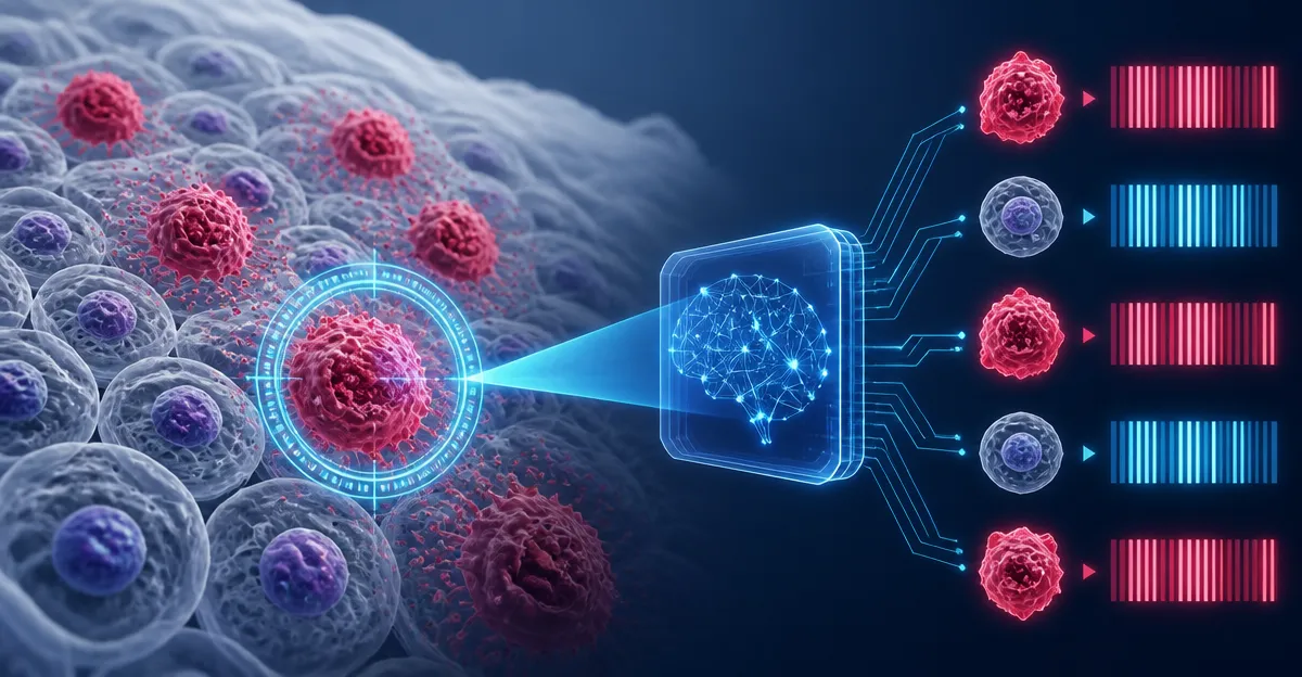
September 21, 2026, (Inside AI) — Researchers at the Massachusetts Institute of Technology have developed a noninvasive method to identify senescent cells, often called "zombie cells," by combining Raman microscopy with single-cell gene expression analysis. The technique, detailed in a study published today in Nature Aging, produces a unique biochemical "barcode" that can flag these cells without destroying them, a crucial step toward diagnosing and treating age-related diseases.
Senescent cells stop dividing but resist dying, accumulating in tissues as we age. They contribute to inflammation, tissue degeneration, cancer, and conditions like osteoarthritis and type 2 diabetes. The immune system normally clears them, but its efficiency wanes over time. Existing biomarkers, such as proteins p16 and p21, require cell-destroying assays, limiting their clinical use.
The MIT team, part of the National Institutes of Health's Cellular Senescence Network, sought a nondestructive alternative. They turned to Raman microscopy, which shines near-infrared or visible light on cells and analyzes the scattered light to reveal chemical composition. By pairing it with spatial RNA sequencing, which maps gene activity within tissue, they examined skin and lung tissue from 2-month-old and 26-month-old mice.
"You can imagine that one day we may develop an endoscope that can look inside your body and identify cellular senescence," says Jeon Woong Kang, an MIT research scientist and one of the senior authors of the study.
Read: GPN-Star Genomic Language Model
The dual approach uncovered a striking increase in lipid synthesis and accumulation in older cells across both tissues. It also revealed tissue-specific changes: senescent skin cells showed altered pathways for muscle contraction and collagen remodeling, while aged lung tissue had heightened immune and inflammatory gene activity. These findings, the researchers say, paint a more comprehensive picture of senescence than either method alone.
"Our idea was to look at many different features to characterize senescence. That's why we wanted to combine both single-cell gene expression and Raman microscopy, so that we can characterize the senescence from two complementary views," says Jian Shu, an assistant professor at Massachusetts General Hospital and Harvard Medical School, and an associate member of the Broad Institute and Ragon Institute.
From the data, the team identified specific Raman peaks—spectral signatures of chemical bonds—that correlate with senescence. These peaks, linked to lipids, proteins, and other molecules, form a barcode that can identify senescent cells in an unbiased way.
"Combining the most important Raman features with the most important gene signatures, we were able to create a barcode that can help us to identify senescent cells in a more unbiased way," says Salvatore Sorrentino, a postdoc at MIT and lead author. "Using this barcode, we can focus on a few Raman bands that emerged as the most informative in this work."
The current system takes about 30 hours to analyze a 1-square-millimeter tissue sample, but the researchers are developing a higher-speed version to scan larger areas quickly. They are also adapting the method for human tissue, though challenges remain in translating findings from mouse models.
"Senescence is not just a pathological condition," says Peter So, director of the MIT Laser Biomedical Research Center and a professor of biological engineering and mechanical engineering. "The idea behind the NIH Cellular Senescence Network is to take a very comprehensive approach to understand senescence and identify senescent cells, because it plays a role in so many normal physiological conditions and many pathological conditions."
The work, funded by the National Institutes of Health and Massachusetts General Hospital, could eventually lead to diagnostics that detect senescent cells during routine endoscopies, enabling earlier intervention. Other groups are exploring senolytic drugs that selectively kill these cells, but without a reliable detection method, their efficacy is hard to gauge. This barcode approach might provide the missing tool.
Read: MIT's GeoPT Teaches AI Physics
As the population ages, therapies targeting senescence could become a cornerstone of preventive medicine. The MIT team's next steps include refining the Raman barcode for human use and speeding up imaging. If successful, the technique might one day be used not only in research but also in clinics to monitor treatments that clear zombie cells.